斑秃(5)-晨读报告
2017年11月20日 0人阅读 返回文章列表
keywords: anagen, catagen, telogen.
–––––––––––––––––––
Late stage
In the late stage of the disease, the inflammation decreases and numerous miniaturized hair follicles and telogen follicles are present. The number of miniaturized follicles increases with chronicity and these may simulate hair follicles in late anagen stage. Such hairs are found in the middle or upper dermis and have been described as nanogen. They represent an intermediate stage between vellus and terminal anagen hair follicles. In horizontal sections there is generally no hair shaft production although occasionally a very thin incompletely keratinized form is produced, correlating with empty infundibula seen on the scalp (Fig. 22.72). In vertical sections the proximal end of the hair shaft acquires a ragged appearance rather than the normal club shape. They can sometimes display histological features of anagen, catagen, and telogen phases simultaneously with evidence of growth and involution in the form of mitotic activity and apoptosis.
晚期(斑秃)
在疾病晚期,炎症减轻,出现大量微小化毛囊和休止期毛囊。微小化毛囊的数量逐渐增加,可与生长末期的毛囊相似。这样的毛发出现在真皮中上部,被成为nanogen,他们代表了介于毳毛和终毛生长期毛囊之间的中间阶段。在水平切片中,一般是没有毛干的,但偶尔会有一个非常细的角化不完全的毛干结构,可在头皮上见到空的漏斗部(图22.72)。在垂直切片中,毛干末梢近端呈锯齿状外观而不是正常的球形。组织学上有时表现为生长期、退行期和休止期同时存在,既有有丝分裂活性,又有凋亡的改变。

Fig. 22.72 Alopecia areata, nanogen hair: the hair shaft shows a decrease in the thickness of the epithelial component with fusion of the internal and external root sheaths. In place of a hair shaft, detritus of amorphous keratin is present.
图22.72 斑秃,nanogen发:毛干显示上皮厚度减少,内外毛根鞘融合。毛干处,存在无定形角质物的碎屑。
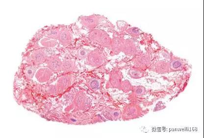
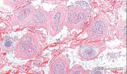
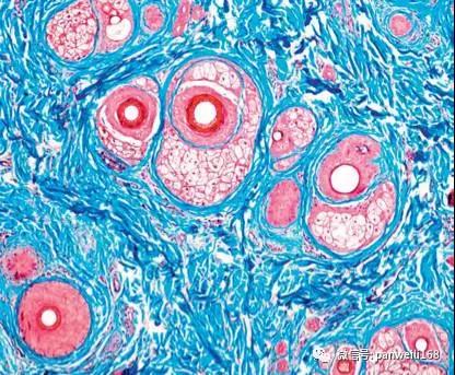
In longstanding alopecia areata the majority of the hair follicles are in catagen and telogen. Since the inflammatory infiltrate does not affect follicles in these growth phases, inflammation may be absent in the subcutaneous tissue (Fig. 22.73).73,84 Inactive alopecia areata can resemble androgenetic alopecia with many miniaturized terminal follicles (Fig. 22.74).
在持续时间长的斑秃中,大多数毛囊处于退行期和休止期。在这两个生长阶段,由于炎症浸润不累及毛囊,所以皮下组织可能没有炎症存在(图22.73)。稳定期斑秃可类似于雄激素源性脱发,有很多微小化毛囊(图22.74)。


Fig. 22.73 Alopecia areata, late stage (A, B): there is a remarkable increase in catagen/telogen follicles, and a sparse peribulbar lymphocytic infiltrate with stellae is evident.
图22.73 斑秃,晚期(A、B):退行期和休止期毛囊显著增加,毛球周围散在的淋巴细胞浸润伴毛囊索很明显。
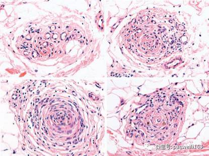
Numerous stellae are present in the deep dermis and the subcutaneous tissue and these may be accompanied by an inflammatory cell infiltrate and melanin pigment(Fig. 22.75). In some cases there may be destruction of the hair follicle by the aforementioned infiltrate and histiocytes and giant cells (Fig. 22.76).
大量毛囊索存在于真皮深部和皮下组织,可能伴有炎症细胞浸润和黑素沉着(图22.75)。在一些病例中,可能出现因上述浸润导致的毛囊破坏以及组织细胞和多核巨细胞(图22.76)。

Fig. 22.74 Alopecia areata, late stage: the similarity to androgenetic alopecia is striking due to the extensive miniaturization and absence of an inflammatory cell infiltrate.Masson's trichrome stain.
图22.74 斑秃,晚期:类似于雄激素源性脱发,因广泛的毛囊微小化且缺乏炎症细胞浸润。Masson三色法。

Fig. 22.75
Alopecia areata: different views of follicular stellae infiltrated by lymphocytes.
图22.75 斑秃:淋巴细胞浸润毛囊索的不同视野。
–––––––––––––––––––––––––––––––
未完待续 (浙江省人民医院皮肤科 陆威 译)2017-11-16

 浙公网安备
33010902000463号


